4shared

Key Benefits
  • Simultaneous stereoscopic 3D images
  • ONH and macula imaging in one click
  • Small pupil mode available
  • 9 point internal fixation for auto mosaic
  • 3D binocular view of the optic cup and disc
  • Confidently diagnose glaucoma patients
  • Fast indentification of disease progression with Kowa’s VK-2 WX3D analysis software 
Specifications
  • Field angle: 45° (Normal mode), 34° (Stereo mode)
  • Working distance 30mm (distance of examined eye to front end of objective lens)
  • Minimum pupil diameter ø4mm (small pupil mode: ø3.5mm)
  • Focusing: Split luminous bars coincidence
  • Working distance adjustment: 2 luminous dots indication type (anterior segment / fundus electrically switched)
  • Internal fixation target: 4 fixed dots switching type
  • Monitor: 5.5 inch LCD monitor
  • Auto OFF function With time setting function
  • Chin rest adjustment range 55mm (electric)
  • Applicable digital camera Specific Nikon digital SLR camera
  • Power Supply Input: AC100V – AC230V 50/60Hz
  • Power Consumption: 200VA (normal) / 250VA (maximum)
  • Dimensions: 310 (W) x 504 (D) x 548 (H) mm
  • Weight: 21kg / 46lbs (excluding the attached digital SLR camera) 
Optional Features
  • External fixation target: Red light, blinking 


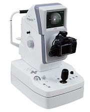
KOWA Nonmyd WX 3D

photo_nonmyd_wx


The Nonmyd WX3D is a versatile retinal camera offering both 2D and superior stereo 3D images while maintaining Kowa's longstanding tradition of high quality imaging and ease of use. The incredibly detailed stereoscopic 3D images delivered through the 3D mode will help you diagnose your patients by providing imaging capabilities unmatched by any other combination retinal imaging device. The Nonmyd WX3Denables 3D viewing of images taken of the ONH and macula, thus providing superior stereo images to support diagnosis of sight threatening conditions such as glaucoma.


Kowa-Ophthalmic-Diagnostics-Nonmyd-WX-3D-Screenshot-05


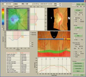
VK2 WX 3D analysis:

  • Depth distribution; Color-coded display of the depth distribution in the analysis area, or graphical display of the cross section of an arbitrary position.
  • 3D display; Display of 3D image based on stereographic data.
  • Numerical data of analusis results; Display of optic disc parameters including "DDLS".
  • Polar coordinates display; The polar coordinates display of the depth distribution permits visual display of the thin part of the rim. (marginal region of cup and disc)